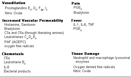
I. The important mediators of acute inflammation

The important substances mediating the processes of acute inflammation are displayed to the right:


II. Mediator Systems

The mediators of acute inflammation are classified into systems based on their source and/or chemical composition.

A. Vasoactive Amines

Histamine, stored in granules in mast cells and released upon stimulation, accounts for the arteriolar dilatation and increased permeability of venules in the immediate transient phase. Histamine is also chemotactic for eosinophils. Other products released by mast cells include PGD2, leukotrienes and AGEPC.
Mast cells are initially stimulated to release their products by trauma, by intracellular components of damaged cells, or by IgE immune complexes. The latter initiates allergic reactions and asthma. Mast cells are also stimulated by activated complement components (C3a, C5a) which may cause anaphylactic reactions in the most severe cases.

Serotonin and histamine are released from platelets upon activation. Platelets are initially activated by contact with collagen and consequent adhesion and also by acetylglycerol ether phosphocholine (AGEPC; also referred to as platelet activating factor - PAF). Though serotonin has the same effects as histamine, vasodilation and increased permeability of venules, in experimental studies with rodents, its action in humans is not established.

The severity of the initial injury determines the degree of mast cell stimulation and the extent of exudation and amount of plasma protein in the exudate. Some of these plasma proteins include promediators which are activated in the interstitial exudate. The C3a and C5a split components of the complement cascade cause degranulation of mast cells during the delayed stage, as do neutrophil lysosomal cationic proteins, thus causing histamine's participation in prolonging exudation. Histamine is inactivated within an hour after its release by histiminase thus preventing prolonged and unbridled effects.

B. Plasma Protein Systems
Increased vascular permeability allows exudation of plasma proteins which include a number of promediator systems. Several mediators are produced as a result of activation of Hageman factor (factor XII --> XIIa) from contact with collagen as plasma proteins effuse through the basement membrane and interstitial matrix. Factor XIIa initiates activation of the Kinin, the Complement and the Clotting/Fibrinolysis systems.

1. The Kinin System
The ultimate product of the kinin system is bradykinin, whose direct effects are:

� smooth muscle contraction
� arteriolar dilatation
� increased permeability of venules (very potent)
� pain

Bradykinin is inactivated by kininases.


2. The Clotting/Fibrinolytic System
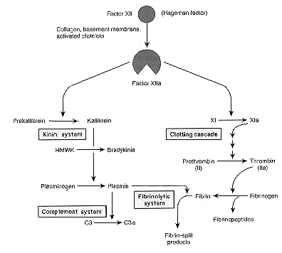
Clotting does not occur until later in the acute inflammatory response unless injury is severe or vasculature is damaged. Early clotting is apparently inhibited by heparin released from mast cells and much fibrinogen (MW = 340,000) probably does not enter the exudate unless vascular permeability is greatly increased from more severe injury. Mediators of the clotting/fibrinolysis system include:

  • thrombin
  • - converts fibrinogen to fibrin and fibrinopeptides
  • fibrinopeptides
  • - induce increased vascular permeability
    - chemotactic for leukocytes
  • plasmin
  • - activates complement components C3 and C5
    - splits fibrin to fibrin split products
  • fibrin split products
  • - may induce increased vascular permeability

    Plasmin is the most important inflammatory mediator of the clotting/fibrinolytic system because of its action in activating C3 and C5 complement components.
    3. The Complement System
    The ultimate objective of the complement system is the formation of the terminal C5-9 assemblage on plasma membranes resulting in the production of large pores effecting cell lysis. Other split products generated as the sequence proceeds are important inflammatory mediators:
    • C3b* - a very short lived form of the C3b fragment which can bind to cell membranes and is then an opsonin for neutrophils and macrophages; otherwise C3b is a component of the C5 convertase and of the alternate pathway C3 convertase
    • C3a - an anaphylatoxin causing mast cell degranulation
    • C5a - an anaphylatoxin causing mast cell degranulation and a chemotactic agent for neutrophils and macrophages, activates phospholipase

    Complement Activation
    Generation of C3b by proteolytic cleavage of native C3 is the critical step for initiating the complement cascade. C3b generation may be initiated by the classical pathway or the alternate pathway.
    • Classic Pathway - initiated by IgM or IgG immune complexes; IgM immune complexes are more effective complement activators. C4b2a is the C3 convertase (and C4b2a3b, the C5 convertase) of the classical pathway. C3 (and C5) may also be converted to their activated products by proteolytic enzymes (plasmin, thrombin, leukocyte lysosomal proteases). Regardless of the mechanism of C3b generation, its production is amplified by the alternate pathway.
    • Alternate Pathway - C3b, protein factors B and D, and Properdin are the constituents of the alternative pathway which results in the production of C3bBb, the C3 convertase of the alternate pathway (and C3bBb.C3b is the C5 convertase).

    C. Prostaglandins and Leukotrienes (Eicosinoids)
    The immediate precursor of the eicosinoids is arachidonic acid, present in the beta position of phospholipids in cell membranes and membranes of subcellular particles. Arachidonic acid is hydrolyzed from phospholipids by Phospholipase A2, also a membrane component. Phospholipase A2 is activated by membrane damage and by C5a.

    Eicosinoids are synthesized via two major pathways:
    • The cyclooxygenase pathway, from which thromboxane A2, prostacyclin (PGI2), and PGD2, PGE2 and PGF2 are synthesized.
    • The 5-lipoxygenase pathway, from which the leukotrienes (LTB4, LTC4, LTD4 and LTE4) are synthesized.

    Aspirin and other NSAIDs inhibit cyclooxygenase but not lipoxygenase. Anti-inflammatory steroids stabilize membranes and prevent phospholipid hydrolysis thereby inhibiting both pathways.
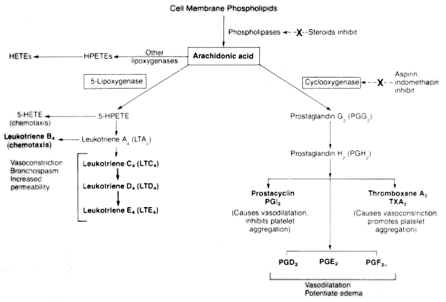
    Different cells have different enzyme systems and synthesize different eicosinoids. Leukocytes, mast cells and platelets synthesize leukotrienes. Platelets also synthesize thromboxane A2 (TXA2). Vascular endothelia synthesize prostacyclin (PGI2). PGD2 is the principal PG synthesized by mast cells. PGD2 and PGE2 are synthesized by a number of different cells.

    Effects of Eicosinoids:

      Prostaglandins:
    • PGE2 -- vasodilates, intensifies bradykinin induced pain, induces fever
    • PGD2 -- vasodilates
    • PGI2  -- vasodilates and is a powerful inhibitor of platelet aggregation
    • TXA2  -- vasoconstricts and stimulates platelet aggregation

      Leukotrienes:
    • LTB4
      - a potent neutrophil and macrophage chemotactic agent
      - causes aggregation of and increased adherence of PMNs to vascular endothelia
    • LTC4, LTD4, LTE4 (identified as the SRS-A)
      - constrict extravascular smooth muscle
      - vasoconstrict
      - increase vascular permeability

    D. Acetyl Glycerol Ether Phosphocholine (PAF)
    AGEPC is a recently identified inflammatory mediator whose effect of activating platelets (platelet activating factor - PAF) has been recognized for some time. AGEPC appears to increase vascular permeability directly as well as from the effect of histamine and serotonin released from platelet activation. AGEPC also causes chemotaxis and leukocyte aggregation and adhesion. AGEPC is synthesized and elaborated by a variety of activated cells including mast cells, phagocytes and endothelia.
    AGEPC also stimulates target cells to synthesize eicosinoids and augments their effects.
    E. Cytokines
    Polypeptide products of activated cells having effects on local and/or distant target cells are referred to as "-kines". Lymphokines are elaborated by activated lymphocytes. Monokines are products of monocytes. Cytokines are elaborated by a variety of cells including lymphocytes, monocytes and endothelial cells. Interleukin 1 (IL-1) and tumor necrosis factor (TNF) are two cytokines implicated to be important inflammatory mediators. IL-1 and TNF cause the same effects in acute inflammation. The actions of the two cytokines include local effects on endothelia, inducing the systemic acute phase reactions and effects on fibroblasts. Interleukin 6 also induces the acute phase response.

    The acute phase reactions effected include:
    � fever
    � sleepiness
    � decreased appetite
    � hemodynamic effects - hypotension, decreased vascular resistance, increased heart rate
    � leukocytosis
    � induction of hepatic synthesis of acute phase proteins (fibrinogen, complement components, C reactive protein)

    The effects on endothelia include:
    � increased synthesis of surface adhesion molecules
    � increased adhesion of leukocytes to endothelium
    � increased elaboration of PGI2 and AGEPC
    � increased thrombogenicity of the endothelial surface

    The effects on fibroblasts include:
    � increased proliferation and collagen synthesis
    � increased collagenase and protease synthesis
    � increased synthesis of PGE2

    IL-8 is a potent chemotactic agent and activator of neutrophils.

    F. Phagocyte Products
    Lysosomal constituents often leak, following phagocytosis, from incompletely enclosed phagocytic vacuoles into the region of inflammation. Lysosomes contain a variety of active proteins. Those which are important in inflammation include cationic proteins and neutral proteases. The heterogeneous group of cationic proteins reside in the azurophil granules of neutrophils and when released have the effects of activating mast cells, of chemotactic activity for monocytes, and of inhibiting the movement of neutrophils and monocytes. Neutral proteases are present in the lysosomes of both neutrophils and monocytes. Unlike accompanying acid proteases which can hydrolyze protein only within the acidic environment of the phagocytic vacuole, the heterogeneous group of neutral proteases exhibit inflammatory activity by hydrolysing a variety of proteins within the site of inflammation including collagen, basement membrane, elastin and cartilage to produce tissue damage as well as fibrin, plasminogen and kininogen.
    The effects of the released lysosomal consituents are prevented from spreading beyond the site of inflammation by the presence in the circulation of inhibitors including alpha-1-antitrypsin and alpha-2-macroglobulin.

    The formation of reactive oxygen metabolites by phagocytes is stimulated by phagocytosis. A portion of the reactive metabolites diffuse into the inflammatory site potentially causing the following effects:
    � endothelial cell damage with consequent increased vascular permeability
    � injury to other cells
    � inactivation of antiproteases

    The significance of the effects of reactive oxygen metabolites during inflammation depends upon the relative rates of formation and of elimination by the antioxidant protective mechanisms: transferrin and ceruloplasmin, superoxide dismutase, catalase and glutathione peroxidase.


    G. Nitric Oxide
    Nitric oxide is a soluble, free radical gas synthesized by vascular endothelia, macrophages, and certain brain neurons. The major effect of NO is vasodilitation. NO also reduces platelet aggregration and adhesion. NO is a free radical and when elaborated by macrophages acts as a cytotoxic agent to attacked microorganisms and neoplastic cells and to tissue in the vicinity. The short life time of NO keeps its effects localized.