Circulating Lipids & Lipoproteins
The major serum lipids include free (unesterified) fatty acids, lysophospholipids, triglycerides, cholesterol (70% esterified with fatty acids), and phospholipids. Lipids are insoluble in aqueous solution and are transported in plasma complexed with specific proteins or apoproteins. Free fatty acids and lysophospholipids are bound by albumin; triglycerides, cholesterol, cholesterol esters and phospholipids exist as large lipid-protein complexes called lipoproteins.
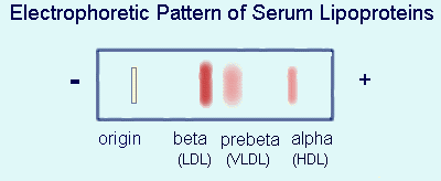 In normal fasting serum, there are three major classes of lipoproteins, which may be resolved by ultracentrifugation or electrophoresis. The lipoprotein classes are interchangibly designated on the basis of their electrophoretic (or corresponding ultracentrifugal) behavior as beta- (or LDL, low density), prebeta- (or VLDL, very low density) and alpha (or HDL, high density) lipoproteins.
For approximately two to eight hours following a meal there is an additional lipoprotein class, called chylomicrons, representing the transport of dietary fat absorbed in the intestine.
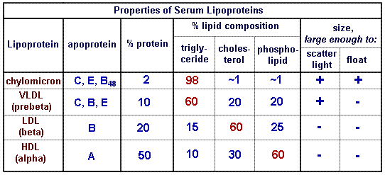
Pertinent properties of the lipoprotein classes are shown in the table below:
Clinical Significance of Serum Lipid Test Results
Elevations of Total Serum Cholesterol and/or Triglyceride
- Lipid elevations are a consequence of one or a combination of the following:
- dietary excess of fat and carbohydrate
- genetic defects in lipoprotein metabolism
- secondary to other disease.
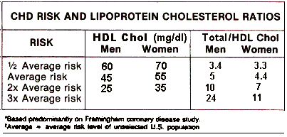
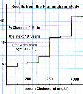 Results from the Framingham Study, a summary of which is depicted to the right, demonstrated a significant relationship between serum cholesterol concentration and the occurrence of MI. Individuals with serum cholesterol concentrations within the upper half of the "normal range" are at a two - three fold greater risk of experiencing a myocardial or cerebral infarct as a consequence of serious atherosclerosis, than are individuals with serum cholesterol concentrations less than 225 mg/dl. Similar epidemiologic studies demonstrated that serum triglyceride concentration was also a risk factor for atherogenisis, though less significant than serum cholesterol. Conclusions from other, more recent, epidemiologic studies are in general agreement with the Framingham study.
Hyperlipidemia from genetic abnormalities in lipid metabolism is more marked and associated with a correspondingly higher risk of disease from atherosclerosis.
_______________________________________________
HDL and LDL cholesterol:
Epidemiologic studies have also demonstrated that decreased HDL is associated with increased incidence of disease from atherosclerosis and increased HDL with decreased atherogenesis. In addition, many of the groups which are known to be at decreased risk (e.g., females, joggers, 2 cocktails/day, etc.) have increased HDL, whereas high risk groups (e.g., diabetics, smokers, etc.) have decreased HDL. It has been proposed that the protective effect of HDL is due to its metabolic role of removing and esterifying free cholesterol from tissues and from the circulation and ultimately presenting it to the liver, via transfer to other lipoproteins, for degradation and excretion into the bile.
Since LDL and HDL are independent risk factors, it is not surprising that their ratio serves as a single risk factor taking into account their combined, and opposite, effects.
Since most of total cholesterol is represented by LDL, the total/HDL cholesterol ratio is considered just as good a risk factor as the LDL/HDL ratio.
Complete genetic deficiency of HDL (Tangier's disease) is associated with a very high risk for disease from atherosclerosis. Heterozygous, mild HDL deficiency is the metabolic defect in familial type IV hyperlipoproteinemia.
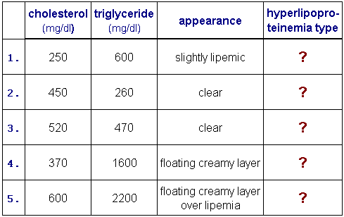
Primary Hyperlipoproteinemia
Primary hyperlipidemia is caused by excessive fat and/or carbohydrate ingestion and/or genetic defects of lipoprotein metabolism. Diagnosis and treatment of hyperlipidemia requires recognition of the responsible lipoprotein(s). Genetic lipoprotein abnormalities are classified into 5 (pheno) types and are generally readily diagnosed on the basis of the alterations in cholesterol and triglyceride concentrations and the appearance of the specimen. A genetic cause for hyperlipidemia can be established only from a family history.
Lipid profile results, total triglycerides, total cholesterol, HDL cholesterol and LDL cholesterol, reported by the laboratory often include interpretive classification of abnormal results in terms of one of the above hyperlipoproteinemia types. The most commonly found types in practice are IV, IIb and IIa.
Type IV is defined as elevated VLDL and is characterized by elevated triglycerides and normal LDL cholesterol. The specimen is usually lipemic when the triglyceride concentration is greater than about 600 mg/dl and the lipemia remains homogeneously distributed after the specimen stands undisturbed overnight.
The metabolic defect is (partial)HDL deficiency. (Tangier's disease is homozygous HDL deficiency.)
Type IIa is defined as elevated LDL and is characterized by elevated LDL cholesterol and normal triglycerides or triglycerides may be slightly elevated but total cholesterol/triglyceride is greater than 2.
The metabolic defect is deficiency of apoB receptors.
Type IIb is defined as elevated LDL and VLDL and is characterized by elevated LDL cholesterol and triglycerides with total cholesterol/triglyceride about 1 (0.7-1.5). Total triglyceride is generally less than 400 mg/dl.
The metabolic defect is a combination of that of Type IIa and Type IV.
Types I, III and V are rare.
Type I is defined as the presence of chylomicrons without an elevation of VLDL as evidenced by lipemia which after standing overnight forms a floating creamy layer over a clear infranate. Triglyceride is elevated and cholesterol is normal.
The metabolic defect is deficiency of lipoprotein lipase.
Type V is defined as the presence of chylomicrons and an elevation of VLDL as evidenced by lipemia which after standing overnight forms a floating creamy layer over a lipemic infranate. Triglyceride is elevated and cholesterol is normal or only slightly increased.
The metabolic defect is a combination of lipoprotein lipase and (partial)HDL deficiency.
Type III is defined as the presence of chylomicron remnants and/or IDL and is characterized by lipemia with elevated triglycerides and cholesterol and total cholesterol/triglyceride is about 1 (0.7-1.5). Type III can be definitively distinguished from Type IIb only by polyacrylamide (molecular sieving) electrophoresis in which Type III exhibits a single band in the slow VLDL position or by ultracentrifugation in which flotation more rapid than beta is exhibited.
The metabolic defect is defective apoE.
Summary of HyperLipoproteinemia Phenotypes
| Type
| metabolic defect
| LP abnormality
| predominant
hyperlipidemia
| appearance next day |
| I
| deficient lipoprotein lipase
| fasting chylomicrons
| hypertriglyceridemia
| floating creamy layer
over clear serum |
| IIa
| deficient apoB receptors
| increased LDL
| hypercholesterolemia
| clear serum |
| IIb
| deficient apoB receptors
and deficient HDL
| increased LDL
| hypercholesterolemia
hypertriglyceridemia
| clear (if trig < 500 mg/dl)
lipemic (if trig > 600 mg/dl) |
| III
| defective apoE
| increased IDL and
chylomicron remnants
| hypercholesterolemia
hypertriglyceridemia
| lipemic |
| IV
| deficient HDL
| increased VLDL
| hypertriglyceridemia
| clear (if trig < 500 mg/dl)
lipemic (if trig > 600 mg/dl) |
| V
| deficient lipoproteinlipase
and deficient HDL
| fasting chylomicrons
and increased VLDL
| hypertriglyceridemia
| floating creamy layer
over lipemic
|
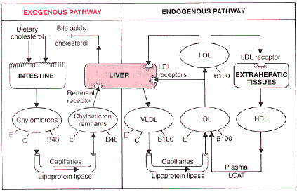 Digested lipids (average dietary intake is about 100 g of triglyceride and 0.5 g of cholesterol per day) are absorbed in the intestinal tract and triglycerides and phospholipids are resynthesized in intestinal epithelial cells. The lipids complex with apoproteins CII, B, and E and are transported in the thoracic lymph duct as chylomicrons. Chylomicrons slowly enter the circulation and are present in blood from about two to about eight hours following a meal. Triglycerides are hydrolyzed by lipoprotein lipase, present on the luminal surface of endothelial cells in muscle and adipose tissue. Triglyceride hydrolysis by lipoprotein lipase requires apoprotein CII, so that extracellular hydrolysis of triglycerides takes place only with chylomicrons and VLDL. Cholesterol and phospholipids from the triglyceride poor chylomicrons are transferred to HDL. The residual chylomicron remnants rapidly interact with hepatocyte membrane receptors for apoproteins B and E and are endocytized and hydrolyzed by lysosomal enzymes.