Properties of Body Fluids

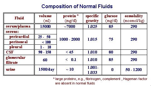
Interstitial fluid and fluid in body cavities is an ultrafiltrate of plasma. The concentration of small molecules, e.g. electrolytes and glucose, is comparable to that in plasma.

The concentration of protein in the fluids is less than that of plasma (~ 6-8 g/dl) and higher molecular weight proteins are excluded to a greater degree, i.e., fibrinogen, alpha-2-macroglobulin, lipoproteins are not present in normal fluids. Membranes lining different cavities vary in their effectiveness in excluding proteins. The protein concentration of normal fluids in the interstitium and in serous cavities is about 1-2 g/dl. The protein concentration of cerebrospinal fluid (CSF) is less than 45 mg/dl. The protein concentration of the glomerular filtrate is less than 0.1 mg/dl.


Hydrodynamic Factors Responsible for Fluid Formation

Starling's Hypothesis for Fluid Formation

The forces responsible for fluid formation are:

 1. Forces in the capillary causing fluid to flow across membranes 
     into the interstitium include:

           capillary hydrostatic pressure (21 mm Hg)
        -  plasma oncotic pressure        (28 mm Hg)
     = - 7 mm Hg

 2. Forces in the interstitium causing fluid to flow back into the capillary include:

           interstitial hydrostatic pressure (1   mm Hg)
         - interstitial oncotic pressure     (8.5 mm Hg)
     = - 7.5 mm Hg

 3. There is a net positive force (~ 0.5 mm Hg) causing the continued formation of 
      extravascular fluid; the excess influx is drained by the lymphatic system. 

Pathologic Serous Fluids

Transudates are fluids of non-inflammatory origin with a normal composition but an increased volume caused by alterations in hydrodynamic factors such as:

Exudates are fluids of inflammatory origin and are characterized by:


Laboratory Tests Conducted on Serous Fluid Collections

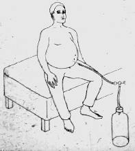
Fluid accumulation causes swelling and/or pain. Symptomatic treatment involves removal of fluid, as depicted in the figure to the right where peritoneal fluid is being aspirated. When necessary, a portion of the fluid is submitted to the laboratory for analysis. Typical laboratory analyses conducted on serous fluids are listed below along with typical results.

  1. visual appearance
    • normal appearance is clear and straw colored with low viscosity.
    • abnormal:
      1. Turbidity indicates a high leukocyte count and, thus, exudation (the extreme case is a purulent exudate). Turbidity is evident when the WBC count is at least 200 cells/ul. The WBC count of a purulent exudate is about 10,000 cells/ul.
      2. high viscosity indicates clotting and/or much hydrolyzed debris
      3. red or pink color indicates the presence of blood (blood in fluid with no history of trauma suggests malignancy)
  2. cytologic examination for malignant cells
  3. culture of microorganisms when infection is suspected
  4. Protein concentration can not always be measured in serous fluids, particularly when viscous, but is estimated from specific gravity measurement
    • normal or transudate: s.g. < 1.015 and protein concentration < 2.0 g/dl
    • ...................exudate: s.g. > 1.018 and protein concentration > 3.0 g/dl
  5. Glucose, likewise, cannot always be measured in viscous fluids, but can be gauged from the following general relationship between protein concentration and leukocyte count and the presumption that leukocytes voraciously consume glucose:
    • normal protein <=> low leukocyte count <=> normal glucose
    • elevated protein <=> high leukocyte count <=> decreased glucose
    Therefore, the glucose concentration is characteristically found to be
    • normal in transudates
    • decreased in exudates, in proportion to the leukocyte count. The glucose concentration is not noticeably decreased unless the WBC count is greater than about 500/ul in which case the fluid will be noticeably turbid.
  6. LDH is increased in exudates from damaged tissue and dead leukocytes. (LDH is the ubiquitous enzyme which catalyzes the last step in glycolysis and its increased presence represents the liberation of intracellular contents into surrounding fluid.) LDH is normal in transudates, except when a lymph duct drains a tumor; the increased LDH results from liberation from rapidly turning over neoplastic tissue.

SUMMARY
Fluid Volume Protein Neutrophils Glucose LDH
Transudate Increased Normal Absent Normal Normal (Increased, if from tumor)
Exudate Increased Increased Increased Decreased Increased


Most Recent Update on 9/2/2014