How variations in TBG concentration cause TT4 results to incorrectly reflect thyroid status can be understood by considering the binding equilibrium:
                                                    Keq
                                   TBG + T4 <-----> TBG-T4

Free and bound T4 are related by:

    free T4 = (bound T4/unoccupied TBG sites)xKeq        (equation 1)

Since greater than 99.9% of total T4 is bound, then bound T4 ~ total T4.  Then,

     free T4 = (total T4/unoccupied TBG sites)xKeq          (equation 2)

Also, since unoccupied TBG sites = total TBG sites - bound T4
        or, unoccupied TBG sites ~ total TBG sites - total T4, then

     free T4 = [total T4/(total TBG - total T4)]xKeq             (equation 3),   or

     free T4 = [(total T4/TBG)/(1 - total T4/TBG)]xKeq        (equation 4)
|

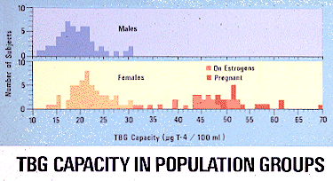
The most common abnormalities of TBG concentration involve approximately 2 fold elevations in pregnancy, in women taking oral contraceptives, or in patients on estrogen therapy for whatever reason, as shown in the figure to the right. Estrogens induce increased hepatic synthesis of a variety of circulating transport proteins, including TBG. On the other hand, decreased TBG concentrations are found in patients with severe, chronic liver disease when hepatic protein synthesizing capacity is compromised and in cases with massive protein loss from the nephrotic syndrome. Congenital abnormalities in TBG synthesis have also been reported.
Determination of TBG, along with total T4, would allow calculation of free T4 by equation 4, above, but direct TBG measurements are not available. The T3 uptake test, described below, does, however, provide an estimate of unoccupied TBG sites and is used in practice to "correct" or "normalize" TT4 concentrations for variations in TBG concentration, by equation 2, above, and provides a value (referred to as the Free Thyroxin Index, FTI) which is proportional to the free thyroxin concentration.
Return to the list of tests in the main document.
T3 Resin Uptake
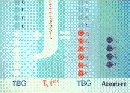
 The principle of the T3 uptake test is illustrated in the figure to the right. The test is conducted by adding a known, excess amount of 125I labeled T3 to a measured volume of serum. The added radiolabeled T3 binds to and saturates the unoccupied TBG sites and does not displace the more tightly bound T4. The excess, unbound 125I T3 is then separated from the serum bound 125I T3 by adding an adsorbent, such as ion exchange resin beads and centrifuging. The serum is decanted, the beads are washed and then counted in a scintillation counter.

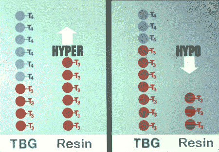
Results were interpreted in the past as illustrated in the figure to the right, assuming normal TBG concentrations. Hyperthyroid cases exhibit a decreased concentration of unoccupied TBG sites and an increased T3 resin uptake. Hypothyroid patients have increased unoccupied TBG sites and decreased T3 resin uptake.
The simpler T3 uptake test was as reliable as the total T4 determination by radioimmunoassay, but is obviously also influenced by variations in TBG concentrations. A euthyroid individual with an elevated TBG concentration would have a low T3 resin uptake and, thus, appear hypothyroid. A euthyroid individual with a low TBG concentration would have a high T3 resin uptake and, thus, appear hyperthyroid, etc. When TBG is abnormal, TT4 and T3RU results indicate opposite conditions. Contrary indications about thyroid status from the two tests, thus, suggest that TBG concentration is abnormal and that each individual result is unreliable.
Return to the list of tests in the main document.
Free Thyroxin Index (FTI, or CT4 or NT4)
The free thyroxin index [ FTI; also referred to as the corrected T4 (CT4) or normalized T4 (NT4) ] utilizes results from both the TT4 determination and the T3 uptake test to provide a value which is proportional to the free thyroxin concentration regardless of TBG concentration. As illustrated in the figures above, the serum uptake of T3* (T3*SU) is proportional to the concentration of unsaturated TBG sites. So that according to equation 2, above,
The FTI provided an accurate indication of thyroid status because it is proportional to the free T4 concentration and is not influenced by variations in TBG concentration as are the individual total T4 and T3 resin uptake determinations.
Return to the list of tests in the main document.
Triiodothyronine (or T3) by RIA
Like T4, total circulating T3 was measured by radioimmunoassay using a specific reagent antibody to T3. Total T3 is influenced by TBG concentration in the same manner as T4 and can likewise by "corrected" with results from the T3 uptake test. The major clinical value in measuring T3 concentrations was that T3 is often more markedly elevated than is T4 in hyperthyroidism. With the advent of the sensitive TSH assay providing this information, T3 by RIA is no longer needed.
Return to the list of tests in the main document.
TRH Stimulation Test
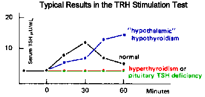
 The TRH stimulation test is used to distinguish between pituitary or hypothalamic deficiency as the cause of secondary hypothyroidisme and to evaluate cases of hyperthyroidism. The test is conducted by collecting blood specimens for TSH before and thirty minutes after intravenous administration of a test dose of TRH. The normal response is a substantial increase in TSH in the 30 minute specimen.
In secondary hypothyroidism caused by pituitary insufficiency the TSH concentration in the thirty minute specimen will remain essentially unchanged. The TSH concentration in the thirty minute specimen will be noticeably increased in cases of hypothalamic insufficiency.
In hyperthyroidism, the pituitary is suppressed by elevated thyroid hormone and there is no significant increase in TSH in the 30 minute specimen.
Return to the list of tests in the main document.
Detection of Thyroid Autoantibodies
Graves' disease, Hashimoto's thyroiditis and primary (autoimmune) myxedema all involve the presence of autoantibodies to thyroid tissue. These auto-antibodies may be directed against microsomal components, thyroglobulin, and/or membrane TSH receptors.
Microsomal and thyroglobulin autoantibodies are determined by most hospital laboratories using latex, or red cell, agglutination methods. These autoantibodies are present in almost all cases of Graves', Hashimoto's and primary (autoimmune) myxedema.
Return to the list of tests in the main document.
Thyroid Scan
Functioning thyroid tissue avidly extracts iodide from the circulation. The rate of iodide uptake depends upon functional activity. Between several hours and a day after the administration of a small dose of 131I, the thyroid is autoradiographed. The autoradiograph may exhibit a diffuse or localized (nodular) pattern of uptake. The major clinical value of the radiologic test is in detecting hyper- or hypofunctioning nodules. A localized "hot" (hyperfunctioning) uptake pattern represents a rare cause of hyperthyroidism and results from the presence of an isolated, hyperfunctioning nodule in a multinodular goiter or of an autonomously functioning adenoma. A "cold" (nonfunctioning) nodule suggests the possibility of carcinoma.
Return to the list of tests in the main document.
Last Updated: March 15, 2000