Pathophysiology of Diabetes Mellitus

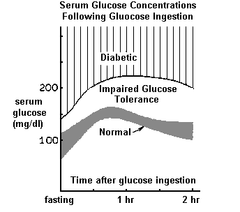
The classic signs and symptoms of diabetes mellitus (hyperglycemia, glycosuria and polyuria, polydipsia, hyperlipidemia, ketonemia and ketonuria, weight loss despite polyphagia) result from inadequate glucose utilization due to insulin lack (Type I) or resistance of tissue to the effect of insulin (Type II). The characteristic time course of serum glucose concentrations following carbohydrate intake is shown below for normal individuals and for diabetics. The immediate consequence of inadequate insulin effect is hyperglycemia from which other signs and symptoms arise.

Glycosuria occurs when the plasma glucose concentration exceeds the renal tubular transport maximum (~ 160 - 180 mg/dl). The presence of significant amounts of glucose in tubule fluid results in polyuria (urine volume > 2 L/day) from osmotic diuresis.

Protein glycosylation, by a spontaneous, nonenzymatic process, is a more recently recognized consequence of hyperglycemia. Glycosylation of proteins in basement membranes is thought to be one factor responsible for the vascular pathology associated with diabetes mellitus. Blood proteins are also glycosylated and remain so during their circulating lifetimes. Determination of glycosylated hemoglobin (Hb A1c), by electrophoresis or column chromatography, provides a means to estimate the extent and duration of hyperglycemic episodes in a retrospective manner and, thus, to monitor the effectiveness of therapeutic control.

Hyperlipidemia is a consequence of increased liberation of fatty acids from adipose tissue because of impaired adipocyte glucose utilization. Triglycerides are continually turned over in adipocytes. Triglyceride synthesis requires a glyerophosphate, the only source of which, in adipocytes, is glycolysis. Lack of a glyerophosphate results in diminished triglyceride resynthesis. Free fatty acids accumulate and are liberated into the circulation at an increased rate and are taken up by the liver and other tissue.
Triglycerides, resynthesized by the liver, are returned to the circulation as prebeta lipoprotein causing Type IV hyperlipoproteinemia secondary to diabetes mellitus.

Keto-acidosis occurs primarily in cases of Type I diabetes and is a consequence of increased utilization of fatty acids as an energy source and increased production of acetylCoA via beta-oxidation. Oxidative phosphorylation is diminished with insulin lack so that acetylCoA accumulates. The excess acetylCoA condenses to acetoacetate and is partially converted to beta hydroxybutyrate and acetone; the three compounds are referred to collectively as ketone bodies. The ketone bodies accumulate in plasma and appear in the glomerular filtrate.


Laboratory Tests

Glucose is measured precisely and accurately in most medical laboratories by highly specific enzymatic methods using hexokinase or glucose oxidase. The enzyme reagents are also impregnated in filter paper strips for use as urine "dipsticks", for patient self monitoring and as a convenient means to measure serum glucose in physicians' offices from finger sticks.
Reagents for detecting ketone bodies are also incorporated into dipsticks for urine testing or into tablets for serum testing.
The term "blood" glucose has persisted (e.g., FBS = fasting blood sugar) since glucose was often measured in whole blood in the past. Whole blood glucose values are less than serum values because the glucose content of erythrocytes is less than that of plasma. Current terminology refers to plasma glucose (e.g., FPG = fasting plasma glucose) eventhough serum is the preferred specimen for analysis. It is best to collect blood specimens in tubes containing fluoride to inhibit glycolysis since erythrocytes metabolize glucose at a rate of about 10 - 20 mg/dl per hour and serum glucose values would otherwise decrease until serum is separated from the clotted red cells.

Screening Tests: Abnormal test result
________________________________________________________________
urine glucose* urine dipstick   - glucose detectable
quantitative   > 25 mg/dl
fasting serum glucose > 110 mg/dl
2 hr. postprandial serum glucose > 140 mg/dl
________________________________________________________________
*The use of urine glucose as a screening test for diabetes is more effective and
sensitive when the urine specimen is collected for several hours following a meal.

Diagnostic Criteria:

Diabetes is not diagnosed on the basis of abnormal screening test results, but on the basis of resultedly abnormal results from testing on fasting, or casual, serum specimens or from the glucose tolerance test.
The glucose tolerance test (GTT) is conducted under highly standardized conditions. The patient is prepared for the test by consuming at least a normal carbohydrate diet (100-150 g/day) for three days prior to the test. On the morning of the test, after a 10-16 hour fast, a blood specimen is collected and if the fasting serum glucose is less than 126 mg/dl, a 75 g test dose of glucose dissolved in water is administered orally within 5 minutes. Blood specimens are collected 1/2, 1, 1-1/2 and 2 hours later and submitted to the laboratory for glucose analysis.
The most recent (February, 2000) diagnostic criteria espoused by the American Diabetes Association are:

  1. Typical symptoms and a casual serum glucose concentration greater than 200 mg/dl, or
  2. fasting serum glucose concentration greater than 126 mg/dl, or
  3. the 2 hour specimen in the oral glucose tolerance test is > 200 mg/dl,
  4. and, upon follow up, a diagnostic value is obtained again from any one of the above tests.
  5. Patients with test results intermediate between normal and definite diabetes are classified as "impaired glucose tolerance" and are not labeled as suspect or latent diabetes, as has been the practice in the past.

Abnormal Glucose Tolerance Secondary to Other Disease

Abnormal glucose tolerance may be secondary to other disease which may need to be evaluated by appropriate laboratory testing. Some conditions recognized to cause abnormal glucose tolerance are listed below:

Pathologic conditions causing impaired, or diabetic, glucose tolerance results: (Normal glucose tolerance is represented by curve 1 and overt diabetes by curve 6.)

  1. Hypercorticism increases the rate of intestinal glucose absorption to produce an early and elevated postprandial peak serum glucose concentration. The additional effects of increased gluconeogenesis and inhibition of glucose uptake in peripheral tissue generally causes an elevated 2 hour postprandial value and an elevated fasting serum glucose. Glucose tolerance results are typified by curve 5.
  2. Acromegaly - Increased growth hormone stimulates glycogenolysis and inhibits the uptake of glucose in peripheral tissue causing elevated fasting and postprandial serum glucose concentrations. Glucose tolerance results are typically between curves 4 and 5.
  3. Hyperthyroidism increases the rate of intestinal glucose absorption to produce an early and elevated peak postprandial serum glucose concentration. The effect of thyroid hormone on peripheral tissue causing increased glucose utilization is generally greater than the effect of increased glycogenolysis on the liver so that the 2 hour postprandial value is usually normal as is the fasting serum glucose concentration. Typical glucose tolerance results are represented by curve 4.
  4. Pheochromacytoma (or "emotional hyperglycemia") - Increased epinephrine increases glycogenolysis resulting in increased fasting and postprandial serum glucose concentrations. Glucose tolerance results are typically between curves 4 and 5.


Pathologic conditions causing flat or depressed glucose tolerance results:

  1. Insulinoma causes rapid uptake of glucose by peripheral tissue resulting in fasting hypoglycemia and a minimal increase in postprandial serum glucose concentrations. Curve 2 or even more depressed glucose tolerance results are typical.
  2. Intestinal malabsorption results in a minimal increase in postprandial serum glucose concentrations. Fasting serum glucose may or may not be subnormal. Glucose tolerance results are typified by curve 2 or even less of an increase of postprandial values.
  3. Low renal Tm for glucose reabsorption results in depressed postprandial serum glucose concentrations and glycosuria even though peak serum values are considerably less than 160 mg/dl. Glucose tolerance results may be normal (curve 1) or may be depressed as in curve 3 if the renal Tm for glucose is sufficiently low.
  4. Hypothyroidism causes a reduced rate of intestinal absorption of glucose and depressed glucose tolerance results typified by curve 2.

Tests for monitoring diabetes

urine glucose and/or ketones
Patient self-monitoring is easily done with urine dipsticks for detecting and semiquantifying glucose and ketones in urine.

glycosylated hemoglobin (HbAlc)
Glycosylated hemoglobin (hemoglobin A1c) determinations are used to ascertain the average, long range effectiveness of therapy. Blood proteins become glycosylated above normal amounts whenever glucose becomes elevated. Increases in glycosylated proteins depend upon the extent, duration and frequency of glucose elevations and the glycosylated proteins remain in the circulation during the proteins' normal lifetimes. Glycosylated hemoglobin is specifically determined because its circulating lifetime is relatively long (about 2 months) and because the glycosylated product is readily distinguished from normal nonglycosylated hemoglobin by electrophoresis or column chromatography.


Diagnosis of Diabetic Coma

Diabetics may become seriously ill or present with life threatening coma requiring prompt therapeutic intervention because of:

  • hypoglycemia from insulin overdose
    Coma may occur when the plasma glucose concentration decreases to values in the vicinity of 25 - 40 mg/dl. Hypoglycemic coma from insulin overdose occurs in an acute manner. Therapy is IV glucose administration.

  • diabetic ketoacidosis
    Type I diabetics are difficult to control even with insulin therapy and are prone to develop ketoacidosis and/or marked hyperglycemia. Serious ketoacidosis develops over a period of hours to days and is associated with marked hyperglycemia (serum glucose is usually in the vicinity of 500 mg/dl or greater) and consequent osmotic diuresis of massive amounts of fluid and electrolyte causes serious dehydration. Serum urea (BUN) and creatinine are elevated about two to four-fold above normal from compensatory decreased renal blood flow. The metabolic, ketoacidosis is associated with a total C02 (bicarbonate) in the vicinity of 10 mEq/L and with an anion gap between 20-30 mEq/L. Maximum possible respiratory compensation maintains the blood pH at about 7.2. Therapy is IV administration of fluid and electrolytes (including bicarbonate) along with insulin.

  • nonketotic hyperosmolality
    Ketoacidosis is rare in non-insulin dependent diabetics. Instead, the non-ketotic hyperosmolar syndrome may be initiated by infection or other physiologic stress. There is serious dehydration from markedly elevated serum glucose, but minimal ketosis and acidosis. The HCO-3 is generally no less than 18 mEq/L.

    Pancreatic Enzymes in Serum for Evaluating Acute Pancreatitis

    Amylase and lipase activity in serum are generally considerably elevated in cases of acute pancreatitis and of obstruction of the pancreatic duct. Mild elevations are found in chronic pancreatitis during periods of exacerbation.

    Serum amylase activity increases as early as six to eight hours following the onset of acute pancreatitis or of duct obstruction and peaks at values 5-10 times the upper limit of normal after about 1 day. Serum activity generally decreases to normal values by three days because of relatively rapid renal clearance (MW=45,000 daltons).
    Urine amylase activity is often elevated earlier than serum activity and remains elevated longer. Urine results are best expressed in terms of clearance with respect to that of creatinine, i.e., fractional amylase clearance = (urine amylase activity x serum creatinine concentration)/(serum amylase activity x urine creatinine concentration). In rare cases, serum activity may be persistently elevated up to about five times the upper limit of normal because of the presence of macroamylases (complexes of amylase with immunoglobulins; the molecular weight of the complex is about 200,000 daltons). Cases of macroamylasemia are revealed by low renal excretion rates. Serum amylase activity is also significantly elevated in inflammatory disease of the salivary glands, however, without an associated increase in serum lipase activity.

    Serum lipase activity peaks to values greater than ten times the upper limit of normal about a day or two later than does serum amylase activity in cases of acute pancreatitis and of duct obstruction and remains elevated for several days longer. Because of its higher molecular weight, lipase is not filtered by the glomeruli as is amylase. Lipase is not elevated in cases of macroamylasemia or in inflammatory disease of the salivary glands.


    Last Updated: January 04, 2001