A. Evaluation of Gastric Acid Secretion
Gastric acid secretion is evaluated in a quantitative manner by measurement of acid in timed collections of gastric contents or in an indirect manner by the Diagnex-blue test. In the Hollander test, gastric acid secretion is measured for the purpose of evaluating therapy for peptic ulcers.
1. Clinical Correlation of Gastric Acid Secretion Rates
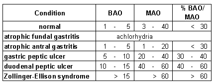 Acid secretion rate is determined from measurement of acid in specimens collected before (BAO) and after (MAO) stimulation (see procedural details). Gastric acid secretion is subnormal or absent (achlorhydria) in cases of chronic fundal gastritis (accompanied by pernicious anemia). Gastric ulceration in the absence of acid output suggests carcinoma since some acid must be present for peptic ulcers to develop.
Acid output is low normal to normal in cases of chronic antral atrophic gastritis. The fundus makes acid but is poorly stimulated because gastrin elaboration by the atrophic antrum is lacking. Gastric peptic ulcers are typically associated with this degree of acid output.
Duodenal peptic ulcer is associated with higher than normal acid secretion
rates.
Gastric acid output is greatest in the Zollinger-Ellison syndrome.
2. The Hollander Test
The purpose of the test is to evaluate the outcome of surgical treatment (gastrectomy and vagotomy) for peptic ulcers.
Acid is measured in gastric fluid collections before and after inducement of hypoglycemia by insulin administration. Hypoglycemia stimulates gastric acid secretion via the vagal nerves and if bilateral vagotomy is complete, MAO increases less than 2 mEq/hr over the BAO.
3. The Diagnex-Blue Test
The test provides an indirect indication of adequate gastric acid secretion
(see procedural details) .
A diagnostic pill, the active ingredient of which is a colored dye bound to a cation exchange resin, is administered orally. The dye is released from the ion exchange resin in the stomach only if there is adequate acid present. Released dye is absorbed by the intestines and is consequently excreted in urine.
The finding of sufficient dye in a urine specimen provides a definite indication of adequate gastric acid secretion and rules out achlorhydria.
Low results are equivocal and direct measurement of acid in gastric fluid collections is required to establish achlorhydria.
B. Serum Gastrin - for diagnosis of the Zollinger-Ellison syndrome.
|
Serum gastrin concentration is determined by radioimmunoassay. Highest serum gastrin concentrations are found in cases of the Zollinger-Ellison syndrome. High serum gastrin concentrations are also found in cases of fundal atrophic gastritis with achlorhydria because of the lack of feedback inhibition. |
| Condition |
serum gastrin
(pg/ml) |
|---|
| normal |
|
| antral gastritis |
low to normal |
| duodenal ulcer |
|
| fundal gastritis |
|
| Z.E. syndrome |
|
|
C. Detection of blood in the stool
The presence of blood in the stool (hematochezia) may result from ulcers or from carcinoma anywhere along the GI tract. If present in sufficient quantity, the blood will be visibly evident as black colored stool from upper GI bleeding (melena) or red colored from lower GI bleeding. "Occult" blood is detectable by laboratory testing using filter paper strips impregnated with guaiac, which turns blue in the presence of peroxidase activity (hemoglobin). About 20 ml of blood must be present to obtain a positive result.
D. The Schilling test - to evaluate vitamin B12 malabsorption.
A consequence of vitamin B12 deficiency is macrocytic anemia and characteristic megaloblastic changes of the myelocyte precursors in bone marrow aspirates. Folate deficiency has the same consequence. Vitamin B12 deficiency is established by finding low serum vitamin B12 concentrations (measured by radioimmunoassay). Vitamin B12 deficiency may be due to pernicious anemia (lack of gastric secretion of intrinsic factor from fundal gastritis) or to intestinal malabsorption or, rarely, to dietary deficiency.
Results from the two stage Schilling test provide a differential diagnosis. (see procedural details) .The test involves evaluation of absorption of two orally administered test doses of radioactive labelled vitamin B12 on the basis of measuring radioactivity in a urine specimen collected for five hours. The first test dose (1st stage) is administered alone and the 2nd test dose (2nd stage, three days later) is administered along with intrinsic factor.
Typical results are shown in the accompanying table.
| With normal vitamin B12 absorption, the test dose will be found to be absorbed, on the basis of finding sufficient radioactivity in the urine specimen, without (1st stage) and with (2nd stage) exogenously administered intrinsic factor. Normal results will be found in cases of vitamin B12 deficiency from chronic dietary lack. In cases of pernicious anemia, malabsorption found in the first stage is ameliorated when intrinsic factor is given in the second stage of the test. In cases of intestinal disease, the test dose is malabsorbed in both stages. |
| Condition |
Results |
| 1st Stage |
2nd Stage |
| normal |
absorption |
absorption |
| dietary deficiency |
absorption |
absorption |
| intestinal disease |
malabsorption |
malabsorption |
| pernicious anemia |
malabsorption |
absorption |
|
E. Tests for steatorrhea
Absorption of dietary fat requires bile salts, pancreatic lipase, formation of bile salt/fatty acid micelles and intestinal absorption. Malabsorption of fat may result from bile duct obstruction, pancreatic in sufficiency
or intestinal disease.
- Indirect indications from decreased absorption of fat soluble vitamins:
- decreased vitamin D absorption ==> negative calcium balance
==> secondary hyperparathyroidism ==> osteomalacia
- decreased vitamin K absorption ==> prolonged prothrombin time ==> susceptibility to hemorrhage
- decreased carotene absorption ==> decreased serum levels determined from O.D. measurement of organic solvent extract
- Microscopic examination of neutral fat stained stool smear.
- Quantitative fecal fat determination on a 72 hr. stool specimen with 100 g/day dietary fat:
- - Normal
| | < | 5 g/day
| |
- - Equivocal
| 5 | - | 7 | |
- - Steatorrhea
| | > | 7 | |
Note: Total fat (including both triglyceride and free fatty acid) is measured, so that steatorrhea from intestinal or pancreaic origin is not distinguished. Determination of the fatty acid/triglyceride composition of a stool specimen might conceivably differentiate pancreatic insufficiency and intestinal malabsorption as the cause, but bacterial lipolytic activity precludes the differentiation.
Nevertheless, the quantitative fecal fat determination provides the most definitive criterion for steatorrhea.
- Measurement of absorption of 125I-labeled oleic acid and 131I-labeled triolein
Procedure: A diagnostic 'fat pill' containing both 125I-labeled oleic acid and
131I-labeled triolein is administered orally. (Use of different isotopes for triolein and oleic acid allows measurement of both isotopes in the same sample. Use of the same isotope would require that the tests for triolein and oleic acid uptake be performed on different days.) A blood specimen is drawn 4 hours later and the 125I and 131I are counted simultaneously. 125I radioactivity reflects free fatty acid absorption and 131I radioactivity reflects triglyceride absorption as illustrated in the figure below.
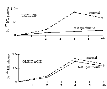
Whereas absorption of triolein is dependent on pancreatic lipase, oleic acid absorption is independent of pancreatic exocrine function. Normal or near normal absorption of labeled oleic acid along with abnormal triolein uptake, as in the figure to the right, suggests impaired pancreatic function.
Failure of absorption of both triolein and oleic acid indicates fat malabsorption of nonpancreatic origin.
F. D-Xlose Test (for evaluation of monosaccharide absorption)
D-xylose is a monosaccharide which is normally readily absorbed by the intestine, is poorly metabolized and is not reabsorbed from the glomerular filtrate.
Following oral administration of a test dose of a solution of the monosaccharide, a blood specimen is collected 2 hours later and a urine specimen is collected for five hours.(see procedural details)
Intestinal malabsorption is reflected in a low amount of D-xylose in the 5 hour urine specimen. (The blood specimen result is important only in cases of renal disease.)
G. Disaccharidase deficiencies
Disaccharidase deficiencies are a cause of colic (intestinal discomfort with vomiting and/or diarrhea) in infants and are not uncommonly acquired in adults.
Lactase or sucrase is most commonly deficient; isomaltase deficiency is rare.
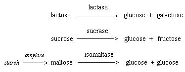
Although the enzyme deficiencies may be evaluated by administration of the disaccharide followed by serum glucose determinations over a two-hour period (tolerance tests) or by assaying biopsy tissue for enzyme activity,
the most practical evaluation is by dietary deletion.
Last updated
on 03/20/2016